Tag Archives: Keratoconus specialists

Keratoconus Specialist in Baltimore

English: Scheme of keratoconus compared to nor...

English: Scheme of keratoconus compared to normal cornea Polski: Schemat porównujący prawidłową rogówkę do stożka rogówki Hrvatski: Skica keratokonusa u usporedbi s normalnom rožnicom (Photo credit: Wikipedia)

Keratoconus is a condition in which the cornea (the lens of the eye) begins to have structural fluctuations, causing it to thin and change to a more conical shape than its normal gradual curve.

The cornea has three major parts, the outer layer, epithelium, the central structural portion, the stroma, which provides the cornea’s shape, and the endothelium, which prevents swelling of the cornea. Keratoconus is a disease of the corneal stroma. The stroma comprises over 85% of the cornea, and is made up of collagen, similar to the material on the tip of your nose. With keratoconus, the cornea loses its usual round shape, and develops in to a cone-like shape.

In the initial stages of keratoconus, vision will fluctuate, causing astigmatism.  As the condition progresses, the cornea becomes too irregular for the use of glasses, and special contact lenses, called Scleral Lenses are needed.  The new generation of Scleral lenses are now very comfortable in the eye and most importantly correct the distortion caused by keratoconus.

Keratoconus is a progressive corneal condition, and regularly starts in the teenage years.  Now with Scleral lenses, people with keratoconus can have great comfortable vision. It’s always in a person’s best interest to avoid corneal transplants and other surgical procedures.

Dr. Irwin Azman specialists in the fitting of Scleral contact lenses for keratoconus, Lasik failures and complications, Pellucid Marginal Degeneration, Stevens-Johnson Syndrome, and other corneal irregularities.
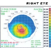
Scleral Contact Lenses for Pellucid Marginal Degeneration

Pellucid Marginal Degeneration:

Pellucid Marginal Degeneration is a sub-category of Keratoconus. Pellucid corneas involve a larger distorted geographic area usually extending from the inferior corneal margins up to the center of the cornea. It is not unusual for 50% or more of the corneal surface to be involved. Because so much of the cornea can be affected, fitting this type of cornea can be challenging. The problem we face as eye care practitioners is fitting the steep areas if the cornea without adversely affecting the flatter areas.

Scleral Lenses:

Scleral contacts are large-diameter gas permeable contact lenses specially designed to vault the entire corneal surface and rest on the “white” of the eye (sclera). In doing so, scleral lenses functionally replace the irregular cornea with a perfectly smooth optical surface to correct vision problems caused by, keratoconus, Lasik failures, post-surgical complications, and other corneal irregularities.

Because scleral lenses are designed to vault the corneal surface and rest on the less sensitive surface of the sclera, these lenses often are more comfortable for a person with corneal irregularities caused by keratoconus and other corneal irregularities  A special liquid fills the space between the back surface of the lens and the front surface of the cornea. This liquid acts as a buffer and protects the compromised corneal tissue. Scleral lenses are designed to fit with little or no lens movement during blinks, making them more stable on the eye, compared with traditional corneal gas permeable lenses. These lenses are almost always very comfortable and the vision provided by them is extremely good. The great majority of patients are able to wear their scleral lenses almost all of their waking hours without problems.

Dr. Irwin Azman, Keratoconus Specialists in Maryland, prescribes scleral contact lenses for a variety of hard-to-fit eyes, including patients with Stevens-Johnson Syndrome, Radial Keratotomy and Lasik Complications and Lasik failures, keratoconus, Corneal Ectasia, Post-Surgical Vision Loss, and Pellucid Marginal Degeneration.

Dr. Irwin Azman specializes in keratoconus, lasik failures and other corneal irregularities neither avoids nor declines the challenge of prescribing the most difficult cases. Dr. Azman tends to have a perfectionist demeanor.

Corneal topogram of a keratoconic eye

Corneal topog. of a keratoconic eye (Photo credit: Wikipedia)