Category Archives: Keratoconus

Keratoconus Specialist: Treating keratoconus with scleral contact lenses, keratoconus glasses, keratoconus hybrid contact lenses, intacts, cross-linking, and other new treatment modalities.

Dr. Oz Show Discusses Long-term Side Effects of LASIK

An episode of the Dr. Oz Show, which aired on October 3rd, 2013, featured an investigative report about the potential life-long side effects of LASIK eye surgery.

As a popular elective surgery, nearly 800,000 people get Lasik every year. FDA clinical data trial shows that every modern laser left 1 out of 5 patients seeing worse after LASIK. Side effects include seeing double, seeing halos or starbursts (especially at night), and dry eye pain.

Investigative reporter Elisabeth Leamy has been investigating the harmful side effects of LASIK for over two years. During her undercover consultations with doctors, she discovered that the side effects were minimalized and the procedure was treated like an easy, no-risk procedure.

Morris Waxler, the former FDA official responsible for getting LASIK surgery approved, is now working to get it banned. He says that the side effects of LASIK are actually injuries and that LASIK harms 20% of patients.

lasik problems

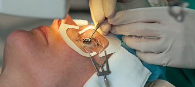
LASIK eye surgery works by cutting a flap of the top layer of the cornea and then using a laser to reshape the exposed cornea. Complications can occur when the flap of the cornea is cut too thick, too thin or too far into the cornea. However, even if this cut is perfect, when the flap is put back down it doesn’t necessarily heal perfectly. Years later it can become dislodged and trigger negative side effects. In addition, if there were underlying issues prior to the LASIK surgery that were not treated, LASIK surgery (and enhancement touch-ups) will most likely exacerbate the issues. Unfortunately many post LASIK patients now have life long side effects, ranging from the smallest amount to the most severe.

Dr. Irwin Azman provides treatment for people who underwent LASIK, PRK, and RK sugery but are now experiencing post-surgical side effects and complications.

According to Dr. Azman:

“While LASIK surgery can be successful under optimum eye conditions, unfortunately many patients do experience side effects due to pre-existing conditions. However, there is hope. Utilizing our vast experience and expertise, as well as an array of new technologies, we are able to treat these side effects with non-surgical techniques.”
For more information about treatments for Post LASIK side effects and complications visit www.lasikfailures.com or call (240) 782-2020.

Diagnosing Keratoconus

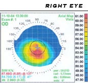
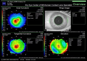
We are now able to detect the presence of keratoconus well before subjective symptoms develop. Diagnosing keratoconus with Wavefront Corneal Topography and Aberrametry measurements have become the standard of care in diagnosing and management of keratoconus. Utilizing new technology, ophthalmic instruments now capture images of the shape of the cornea together with the entire optical system by taking tens of thousands of data points. The results are instantly analyzed and Wavefront topography maps are generated. These printouts will show Dr. Azman a map of the eye-print  (just like a finger print) of the location and severity of any corneal distortion and high order aberrations (HOA).
Keratoconus w/ IntacsKeratoconus Uncorrected with HOAsKeratoconus Corrected with contact lenses
Corneal specialists agree that it is best to exhaust all non-surgical options for keratoconus before undergoing corneal transplant surgery, especially for younger patients. Optometrists and Ophthalmologists from all over Maryland and surrounding states refer their challenging keratoconus patients to Dr. Azman.
Keratoconus Evaluation
Tear Scan
Tear Osmolarity
Anterior OCT
Posterior OCT
Tear Film Analysis and Dry Eye Evaluation
Lid Evaluation
Wave front corneal topography
Aberratometry
Specular Microscopy
Refraction
Pupil measurement
Ulltrasound corneal thickness
Keratoconus Treatment Options
Keratoconus Glasses
Scleral contact lenses
Keratoconus soft contact lenses
Hybrid contact lenses
IPL
LIPIFLOW
CRT CORNEAL Reshaping Therapy
Dry eye treatment
Intacs
Cross linking
Corneal transplant(last option)
Prescription and/or OTC medication
Tear ScanAnterior OCTPosterior OCT

Keratoconus Specialist in Baltimore

English: Scheme of keratoconus compared to nor...

English: Scheme of keratoconus compared to normal cornea Polski: Schemat porównujący prawidłową rogówkę do stożka rogówki Hrvatski: Skica keratokonusa u usporedbi s normalnom rožnicom (Photo credit: Wikipedia)

Keratoconus is a condition in which the cornea (the lens of the eye) begins to have structural fluctuations, causing it to thin and change to a more conical shape than its normal gradual curve.

The cornea has three major parts, the outer layer, epithelium, the central structural portion, the stroma, which provides the cornea’s shape, and the endothelium, which prevents swelling of the cornea. Keratoconus is a disease of the corneal stroma. The stroma comprises over 85% of the cornea, and is made up of collagen, similar to the material on the tip of your nose. With keratoconus, the cornea loses its usual round shape, and develops in to a cone-like shape.

In the initial stages of keratoconus, vision will fluctuate, causing astigmatism.  As the condition progresses, the cornea becomes too irregular for the use of glasses, and special contact lenses, called Scleral Lenses are needed.  The new generation of Scleral lenses are now very comfortable in the eye and most importantly correct the distortion caused by keratoconus.

Keratoconus is a progressive corneal condition, and regularly starts in the teenage years.  Now with Scleral lenses, people with keratoconus can have great comfortable vision. It’s always in a person’s best interest to avoid corneal transplants and other surgical procedures.

Dr. Irwin Azman specialists in the fitting of Scleral contact lenses for keratoconus, Lasik failures and complications, Pellucid Marginal Degeneration, Stevens-Johnson Syndrome, and other corneal irregularities.

Scleral Contact Lenses for Pellucid Marginal Degeneration

Pellucid Marginal Degeneration:

Pellucid Marginal Degeneration is a sub-category of Keratoconus. Pellucid corneas involve a larger distorted geographic area usually extending from the inferior corneal margins up to the center of the cornea. It is not unusual for 50% or more of the corneal surface to be involved. Because so much of the cornea can be affected, fitting this type of cornea can be challenging. The problem we face as eye care practitioners is fitting the steep areas if the cornea without adversely affecting the flatter areas.

Scleral Lenses:

Scleral contacts are large-diameter gas permeable contact lenses specially designed to vault the entire corneal surface and rest on the “white” of the eye (sclera). In doing so, scleral lenses functionally replace the irregular cornea with a perfectly smooth optical surface to correct vision problems caused by, keratoconus, Lasik failures, post-surgical complications, and other corneal irregularities.

Because scleral lenses are designed to vault the corneal surface and rest on the less sensitive surface of the sclera, these lenses often are more comfortable for a person with corneal irregularities caused by keratoconus and other corneal irregularities  A special liquid fills the space between the back surface of the lens and the front surface of the cornea. This liquid acts as a buffer and protects the compromised corneal tissue. Scleral lenses are designed to fit with little or no lens movement during blinks, making them more stable on the eye, compared with traditional corneal gas permeable lenses. These lenses are almost always very comfortable and the vision provided by them is extremely good. The great majority of patients are able to wear their scleral lenses almost all of their waking hours without problems.

Dr. Irwin Azman, Keratoconus Specialists in Maryland, prescribes scleral contact lenses for a variety of hard-to-fit eyes, including patients with Stevens-Johnson Syndrome, Radial Keratotomy and Lasik Complications and Lasik failures, keratoconus, Corneal Ectasia, Post-Surgical Vision Loss, and Pellucid Marginal Degeneration.

Dr. Irwin Azman specializes in keratoconus, lasik failures and other corneal irregularities neither avoids nor declines the challenge of prescribing the most difficult cases. Dr. Azman tends to have a perfectionist demeanor.

Corneal topogram of a keratoconic eye

Corneal topog. of a keratoconic eye (Photo credit: Wikipedia)


Corneal Ectasia: Scleral Lenses

My Cornea looks like a Black hole!

(Photo credit: Rakesh Rocky)

 Corneal Ectasia:

A condition resembling Keratoconus is virtually always caused by refractive eye surgery, specifically LASIK. After LASIK, the cornea has been made thinner. Because the corneal “wall” has been made thinner, internal pressure from within the eye can cause expansion or distension of the cornea. The resultant distorted corneal surface will usually make it impossible to have clear vision with eyeglasses or soft contact lenses. Most of the time a special gas permeable contact or scleral lens will be needed to restore lost vision. These special high tech lenses (which have only recently become available) will act to create a new corneal surface allowing the patient to regain clear, comfortable vision.

Post Lasik Treatments:

There are a number of specialized lenses now available that will allow post-surgical patients to function on a much higher level and with a much greater level of comfort. To address the vision and comfort issues that most post-refractive surgical patients are facing, Dr. Azman prescribes Post-Surgical Scleral Lenses.

Scleral contacts are large-diameter gas permeable contact lenses specially designed to vault over the entire corneal surface and rest on the “white” of the eye (sclera). In doing so, scleral lenses functionally replace the irregular cornea with a perfectly smooth optical surface to correct vision problems caused by Lasik failures, post-surgical complications, and other corneal irregularities.

Because scleral lenses are designed to vault the corneal surface and rest on the less sensitive surface of the sclera, these lenses often are more comfortable for a person with corneal irregularities. A special liquid fills the space between the back surface of the lens and the front surface of the cornea. This liquid acts as a buffer and protects the compromised corneal tissue. Scleral lenses are designed to fit with little or no lens movement during blinks, making them more stable on the eye, compared with traditional corneal gas permeable lenses. These lenses are almost always very comfortable and the vision provided by them is extremely good. The great majority of patients are able to wear their scleral lenses almost all of their waking hours without problems.

Dr. Irwin Azman prescribes scleral contact lenses for a variety of hard-to-fit eyes, including patients with Stevens-Johnson Syndrome, Radial Keratotomy and Lasik Complications, Keratoconus, Corneal Ectasia, Post-Surgical Vision Loss, and Pellucid Marginal Degeneration.

Dr. Irwin Azman neither avoids nor declines the challenge of prescribing the most difficult cases. Dr. Azman tends to have a perfectionist demeanor.


Keratoconus Treatment

Keratoconus:

Dr. Irwin Azman is one of the few contact lens specialists that has extensive experience in the diagnosis and management of the Keratoconus patient. With over 30 years of experience, his team of technicians, new technologies in instrumentation, contact lens materials and designs, Dr. Irwin Azman is able to prescribe each Keratoconus patient with the perfect fit.

Diagnosing Keratoconus:

We are now able to detect the presence of Keratoconus well before subjective symptoms develop. Corneal Topography and Aberrametry measurements have become the standard of care in diagnosing and management of Keratoconus. A computerized system images the shape of the cornea together with the entire optical system by taking tens of thousands of data points. The results are instantly analyzed and wave front topography maps are generated. These printouts will show Dr. Azman a map print (just like a finger print) of the patient’s location and severity of any corneal distortion and high order aberrations (HOA).

Corneal Wavefront Topography

Corneal Wavefront Topography

Azman Contact Lens Specialists is proud to be the first and only ones in the state of Maryland to employ both new technologies, Marco3-D Wave Corneal Analysis and the Opthonix aberrometer for Keratoconus treatment and management.

Call for a complimentary phone consultation! (240) 782-2020

Health/Life Style and Medical History:
Information concerning your medical health status eye medical status, allergies, family history, and life style and sport activities. Many patients that have allergies have Keratoconus.

Corneal Topography:
The computerized corneal topography measures the cornea’s surface combining, wavefront, auto-refraction, keratometry, and pupillometry — allowing accurate and reliable analysis of corneal High and Low order aberrations. For you, the patient, it is a brief, painless, non-contact procedure that photographs your eye’s surface.

Specular Microscopy:
A noninvasive photographic technique that facilitates accurate and precise diagnosis of corneal disease in the back part of the cornea (endothelium). This instrument can pick up early signs of corneal edema, which cannot be picked up on routine examination. If there is any indication of corneal disease, such as Keratoconus or edema we might recommend a different treatment modality.

Keratoconus Treatment: Scleral Lenses

Corneal specialists agree that it is best to exhaust all non-surgical options for Keratoconus before undergoing corneal transplant surgery, especially for younger patients. Optometrists and Ophthalmologists from all over Maryland and surrounding states refer their challenging Keratoconus patients to Dr. Irwin Azman.

Scleral contacts are large-diameter gas permeable contact lenses specially designed to vault over the entire corneal surface and rest on the “white” of the eye (sclera). In doing so, scleral lenses functionally replace the irregular cornea with a perfectly smooth optical surface to correct vision problems caused by Lasik failures, post-surgical complications, and other corneal irregularities.

Because scleral lenses are designed to vault the corneal surface and rest on the less sensitive surface of the sclera, these lenses often are more comfortable for a person with corneal irregularities. A special liquid fills the space between the back surface of the lens and the front surface of the cornea. This liquid acts as a buffer and protects the compromised corneal tissue. Scleral lenses are designed to fit with little or no lens movement during blinks, making them more stable on the eye, compared with traditional corneal gas permeable lenses. These lenses are almost always very comfortable and the vision provided by them is extremely good. The great majority of patients are able to wear their scleral lenses almost all of their waking hours without problems.

Dr. Irwin Azman prescribes scleral contact lenses for a variety of hard-to-fit eyes, including patients with Stevens-Johnson Syndrome, Radial Keratotomy and Lasik Complications, Keratoconus, Corneal Ectasia, Post-Surgical Vision Loss, and Pellucid Marginal Degeneration.

Dr. Irwin Azman neither avoids nor declines the challenge of prescribing the most difficult cases. Dr. Azman tends to have a perfectionist demeanor.